Diagnosing, Treating & Managing IVDD
Diagnosing IVDD
Diagnosis of spinal cord compression caused by IVDD requires imaging of the spinal cord. The gold standard for spinal cord imaging is magnetic resonance imaging (MRI). In some circumstances, computed tomography (CT) can diagnose ruptured mineralized disks with spinal cord compression.
Treating and Managing Dogs with IVDD
There is a diversity of opinion regarding treatment options for dogs with IVDD, but general guidelines can be used for selecting therapy. Decisions regarding when and if surgical versus medical treatment for spinal compressive disease is indicated depend primarily upon the severity of the neurological signs and the chronicity of the problem. In addition, treatment is modified in relation to the presumptive diagnosis, owner finances, and concomitant medical problems. Medical treatment should be recommended only if the clinician has great confidence in a presumptive diagnosis of IVDD
The cornerstone of medical management of IVDD is cage confinement with short, supervised visits outside to urinate and defecate. They should be on leash and harness at all times when they are not in a small crate or in a quiet room with no disturbance. Other components of medical management can include pain medications (gabapentin, narcotics), muscle relaxants, and anti-inflammatories. If your patient improves with medical management, continued treatment is indicated for two weeks after the animal is clinically normal.
If clinical signs are not improved after one or two weeks or the dog worsens, definitive diagnosis and surgery should be considered.
or a client declines definitive diagnosis and treatment.
In the case of an older patient or one that is clinically ill (e.g. anorexia, weight loss, fever, etc.), a more aggressive diagnostic course of action is warranted.
GRADES 3-6: These animals are surgical candidates.
Grade 3 motor dysfunction refers to an animal that still has movement but cannot bear significant weight or propel themselves on their own.
Animals with Grade 4 neurological dysfunction have no motor, are completely paralyzed but still have sensation of deep pain perception (see Fig X(Bailey)). If these animals have surgery performed have an excellent recovery rate (90-95%).
Grade 5 animals (those that have lost the perception of deep pain for less than 48 hours) that are operated on still have a fair to good prognosis for recovery (60-80%).
If an animal has lost deep pain for more than 48 hours (Grade 6), a guarded prognosis should be given to the owner, although recent reviews indicate a 50% recovery rate.
Animals in the Grade 5 and 6 categories may require an emergency myelogram if the preferred imaging modalities of CT or MRI are not available in a timely manner prior to surgery in order to definitively diagnose and localize the ruptured disk.
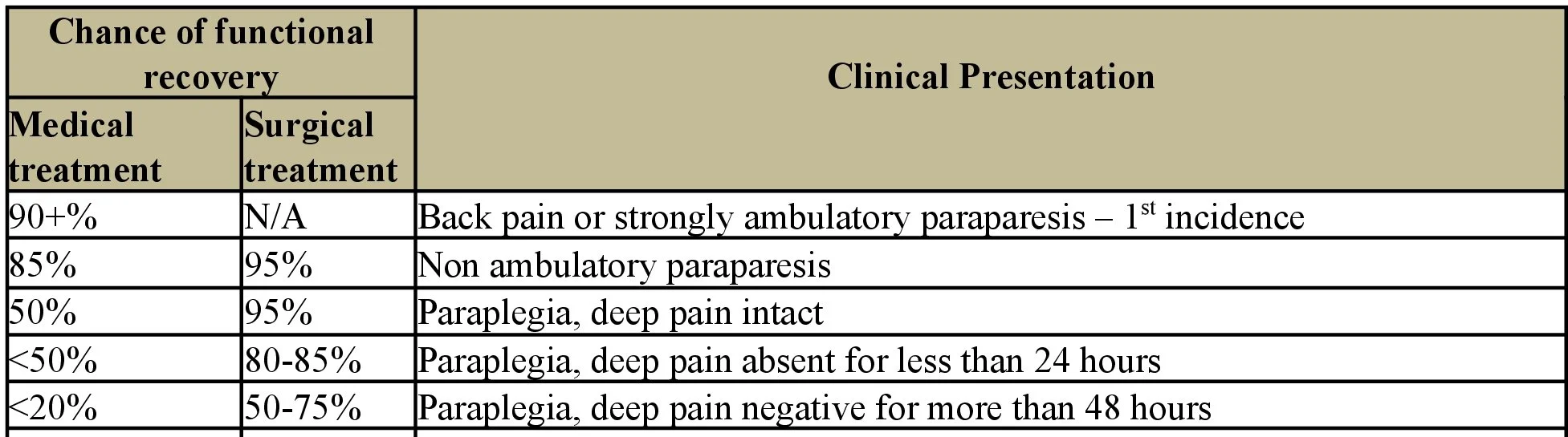
Neuro statistics to remember - Intervertebral disk disease