NEUROLOGICAL DISEASES IN PETS:
Diskospondylitis
Diskospondylitis is a bacterial or fungal infection of the intervertebral discs and adjacent vertebrae. It primarily affects large, middle-aged to older pets, causing pain, stiffness, and lethargy. The infection can spread through the bloodstream or from a nearby wound. Diagnosis involves imaging and lab tests. Treatment includes long-term antibiotics and pain management. Early and aggressive treatment generally leads to a good prognosis, although relapses can occur.
Disease Overview
Diskospondylitis is an infection of the intervertebral disks and adjacent vertebral endplates caused by bacteria or fungi. It primarily affects dogs, particularly those that are large, middle-aged to older, although it can rarely occur in cats. Male dogs are more frequently affected than females.
The signs of this condition are often vague, leading to it being referred to as “the great imitator.” Affected pets may exhibit symptoms such as pain, discomfort, lethargy, reduced appetite (hyporexia), weight loss, and overall poor condition (unthriftiness). Other common signs include stiffness, reluctance to move, muscle weakness, fever, and in severe cases, paralysis.
The infection can occur due to hematogenous spread or directly from a wound. Cases with systemic or hematogenous spread (through the blood stream) can have involvement in the urinary system, eyes, spleen, lymph nodes, or heart valves. Diskospondylitis can also result from direct contamination through a puncture or bite wound near the spine. Additionally, infections may arise in association with a migrating foreign body near the vertebral column. The most common bacterial causes of diskospondylitis include E. coli, Staphylococcus, Streptococcus, and Brucella; fungal organisms can occasionally be responsible, as well.
Diskospondylitis can occur at a single location within the spinal column or at multiple sites. The most commonly affected disks are L7-S1, T13-L1, and C6-7-T1. More than 40% of dogs will have multiple discospondylitis lesions. The signs of diskospondylitis typically begin gradually and worsen over time. In many instances, the first noticeable sign is back pain. Pets may appear stiff, be reluctant to jump on or off furniture or express pain when turning a certain way or when touched in a specific area.
As the condition progresses, additional symptoms may include stiffness and muscle weakness in the limbs. In severe cases, pets may become paralyzed. Many affected animals, although not all, also exhibit nonspecific signs such as decreased appetite, weight loss, and lethargy.
Radiographs can suggest the presence of diskospondylitis; however, radiographic changes may emerge more than two months after clinical symptoms appear. These radiographs can be used to monitor the response to treatment as well.
Diagnosing Diskospondylitis in Dogs and Cats
Advanced imaging techniques, such as computed tomography (CT) and magnetic resonance imaging (MRI), are valuable tools for diagnosing diskospondylitis. Definitive diagnosis of the causative organism can be obtained through surgical exploration, biopsy, or disk aspiration.
Pets diagnosed with diskospondylitis generally require baseline testing, which includes blood work (complete blood count and chemistry panel), urinalysis, and urine culture and sensitivity testing to identify the infectious bacteria and determine the most effective medication. Additional tests may involve blood tests for Brucella and possibly blood cultures or an ultrasound of the heart.
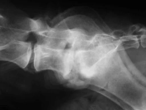
Lateral projection radiograph of the lumbosacral junction of an 8-year-old MN Labrador Retriever that presented with trouble posturing to defecate, pain, and trouble jumping into the car. The radiograph demonstrates an exuberant proliferation of bony reaction at the LS, sclerosis of the endplates, and lysis of the endplates of the LS disk space.
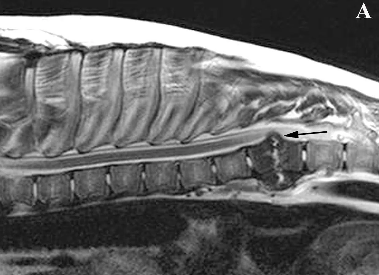
Magnetic Resonance Imaging (MRI) of a male 10-year-old Chesapeake Bay Retriever presenting for back pain and weakness in his back legs. A sagittal T1 W post-contrast image of the thoracic vertebral column and spinal cord is shown. At T8-9, there is dramatic sclerosis of the vertebral bodies, erosions of the vertebral endplates (caudal endplate of T8 and cranial endplate of T9), and contrast-enhancing material within the disk space. The material within the disk space protrudes into the spinal canal, causing severe compression of the spinal cord.
Treatment Options for Diskospondylitis in Dogs and Cats
Occasionally, spinal surgery may be necessary to remove infected tissue and obtain samples for culture and sensitivity testing. In the majority of cases, treatment for diskospondylitis typically involves intravenous (IV) or oral antibiotics, ideally chosen based on culture and sensitivity results. Antibiotic treatment is long-term, usually lasting at least three months, although it can extend longer if needed. Pain medications are also prescribed, as most dogs experience significant pain.
Concerned about Diskospondylitis in your pet? Contact a veterinary neurologist for an accurate diagnosis and effective treatment. Schedule your appointment with us today.
Strict rest is crucial during the early recovery process, as the vertebrae, supporting ligaments, and muscles become weak and are susceptible to injuries, such as fractures and subluxations. These injuries can be severe and challenging to repair if they occur. Close monitoring and follow-up examinations are recommended, and rechecking radiographs every six to eight weeks helps assess whether healing is progressing or if the infection is ongoing.
Prognosis for Diskospondylitis in Dogs and Cats
The prognosis for dogs affected by diskospondylitis depends on the underlying cause of the infection. Bacterial diskospondylitis often has a favorable prognosis. Most cases resolve with early and aggressive treatment, although six to twelve months of antibiotics may be required, and relapses can occur.
Diskospondylitis in Cats
Although rare, Diskospondylitis can affect cats and presents similarly with symptoms such as pain, lethargy, and decreased appetite. Diagnosis and treatment protocols are similar to those for dogs, involving imaging, long-term antibiotics, and pain management. Cats often exhibit subtler symptoms, making early detection critical.
Frequently Asked Questions about Diskospondylitis in Dogs and Cats
-
Diskospondylitis in dogs is typically caused by bacterial or fungal infections. These infections may spread through the bloodstream or result from a nearby wound.
-
No, Diskospondylitis is rare in cats, but when it occurs, it can cause significant discomfort and requires prompt treatment.
-
Treatment usually involves long-term antibiotics based on culture and sensitivity results, pain management, and, in severe cases, surgery to remove infected tissue.
Examples of Diskospondylitis Cases
PATIENT 1: Shiloh
Shiloh’s lateral radiograph of the lumbosacral (LS) junction (arrow) showing collapse and subluxation of the LS, sclerosis and lysis of the endplates at L7 and S1, periosteal reaction along the vertebrae, and spondylosis at the joint.
Similar site by MRI of Shiloh’s LS. This is a T1W post-contrast sagittal view. There is inflammation indicated by contrast-enhancement around the spinal cord, dorsal aspect of the LS disk, within the endplates and surrounding tissue, and the interarcuate ligament.
PATIENT 2: ANGIE
Angie, a 9 ½-year-old female spayed Abyssinian cat presented to us with back pain and inability to jump on furniture. Her examination indicates a problem within the thoracolumbar spine and focal pain in the middle of her thoracic spine.
Angie’s lateral radiograph of her thoracic spine. The arrow points to an area of intervertebral disk collapse, endplate sclerosis and lysis, and bridging spondylosis characteristic for a diskospondylitis lesion at the T9-10 disk space.
Concerned about Diskospondylitis in your pet?
Contact a veterinary neurologist for an accurate diagnosis and effective treatment. Schedule your appointment today.


