NEUROLOGICAL DISEASES IN PETS:
Acute Non-Compressive Nucleus Pulposus Extrusion (ANNPE)
(aka 'high-velocity low volume' disk, Hansen type III IVDD, traumatic disk)

Disease Overview
Acute Non-Compressive Nucleus Pulposus Extrusion (ANNPE) is a condition that leads to acute, asymmetric thoracolumbar myelopathy. Other terms include traumatic disk extrusion, Hansen Type III disk, high-velocity low-volume disk extrusion, and intervertebral disk explosion. ANNPE occurs when a small amount of hydrated disk material explosively extrudes into the spinal canal, disrupting the spinal cord’s vascular supply. According to the American College of Veterinary Internal Medicine (ACVIM), this condition can result in acute spinal cord trauma requiring prompt veterinary attention. This typically happens due to excessive force applied to a healthy disk, often caused by trauma, falls, or vigorous exercise. Although the extruded material minimally compresses the spinal cord, it can still cause direct injury and bruising, leading to sudden spinal cord trauma.
ANNPE can occur in any dog breed but is most common in young, active, medium to large dogs. It frequently affects working, sporting, or high-drive dogs, such as Border Collies, Labrador Retrievers, German Shorthaired Pointers, and Australian Cattle Dogs. The condition is usually not painful and stabilizes within 24-48 hours.
Diagnosing ANNPE in Dogs
Advanced imaging and CSF sampling are essential for diagnosing ANNPE. Magnetic resonance imaging (MRI) is the most definitive diagnostic tool, distinguishing ANNPE from compressive disk disease, spinal cord tumors, and inflammatory conditions. According to the American College of Veterinary Radiology, MRI findings can also help provide a prognosis based on lesion location and spinal cord involvement. CSF sampling may be performed to rule out other diseases that mimic ANNPE. If MRI is unavailable, a presumptive diagnosis can be made through medical history, physical exams, and characteristic recovery patterns.

Sagittal view of patient’s spine - MRI STIR sequence. The red arrow points toward the lesion in the middle of the spinal cord which is bright in this sequence and lies right above the T13-L1 disk space.
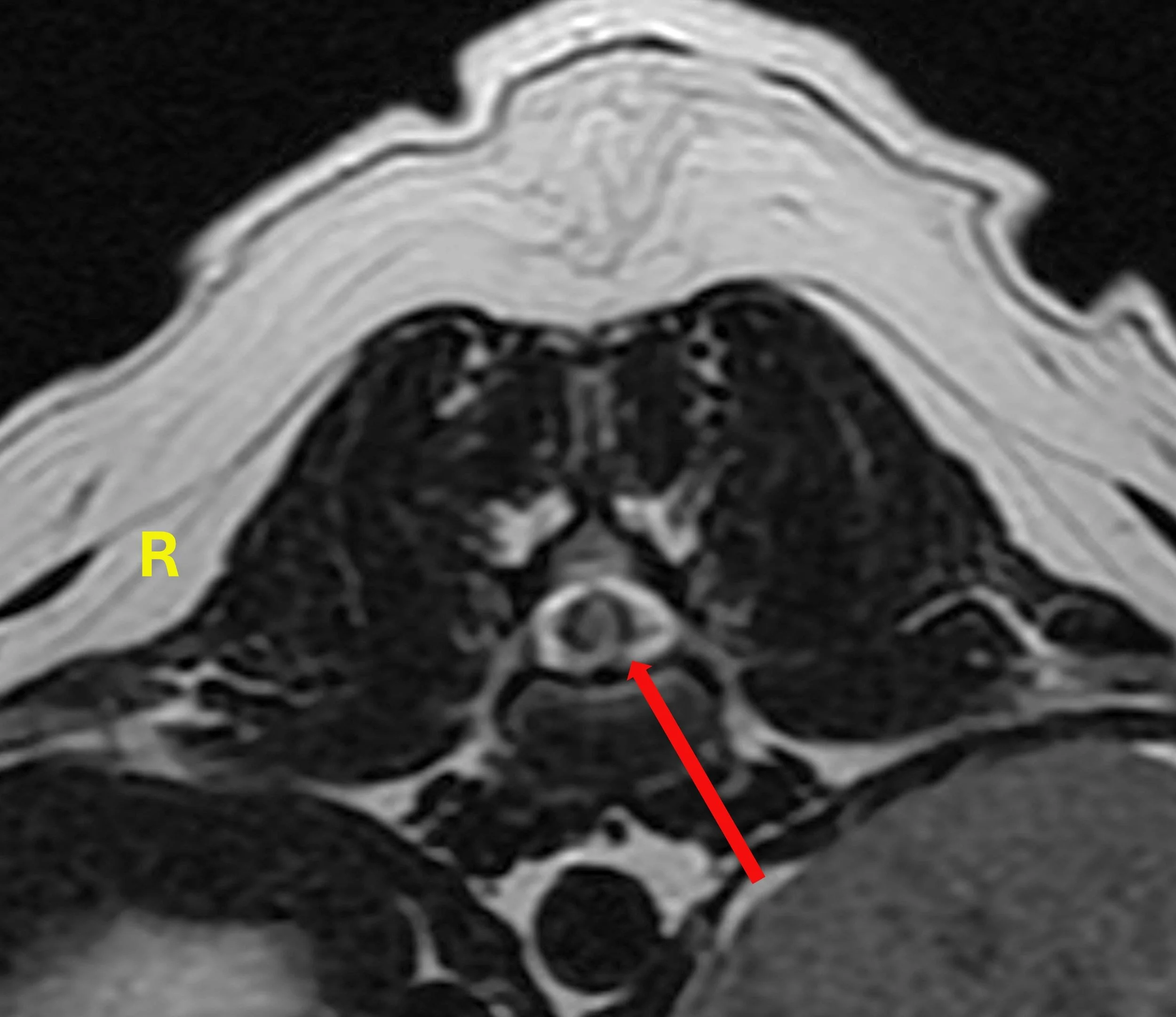
Axial view of patient’s spine at the level of the T13-L1 disk space - MRI T2W sequence. The red arrow points towards the bright spot on the left side of the spinal cord (left and right projections are opposite in imaging figures).
Treatment Options for ANNPE in Dogs
ANNPE treatment depends on severity and includes nursing care, physiotherapy, pain management, and bladder care. Physical activity restrictions for 4-6 weeks may be recommended to allow spinal cord healing. Provide clean, soft bedding to prevent bedsores and assist with changing sides every 4-6 hours if needed. Ensure easy access to food and water, and manually express the bladder if the dog cannot urinate voluntarily. Be vigilant for signs of urinary tract infections due to incomplete bladder emptying. Pain management should be coordinated with a veterinarian as needed.
Physiotherapy and veterinary rehabilitation are crucial for recovery, helping prevent complications such as joint stiffness, muscle atrophy, and pressure sores. According to the American Veterinary Medical Association (AVMA), veterinary-supervised rehabilitation enhances recovery through structured therapies.
Prognosis for ANNPE in Dogs
The prognosis for dogs recovering from ANNPE varies based on the severity of the spinal cord injury. Preservation of deep pain sensation (the ability to detect painful stimuli on the toes) indicates a better chance of recovery. While mild injuries may resolve quickly, severe cases can require months of rehabilitation and may leave residual neurological deficits. Each case is unique, and recovery outcomes vary accordingly.
Juno: An ANNPE Patient Case Study
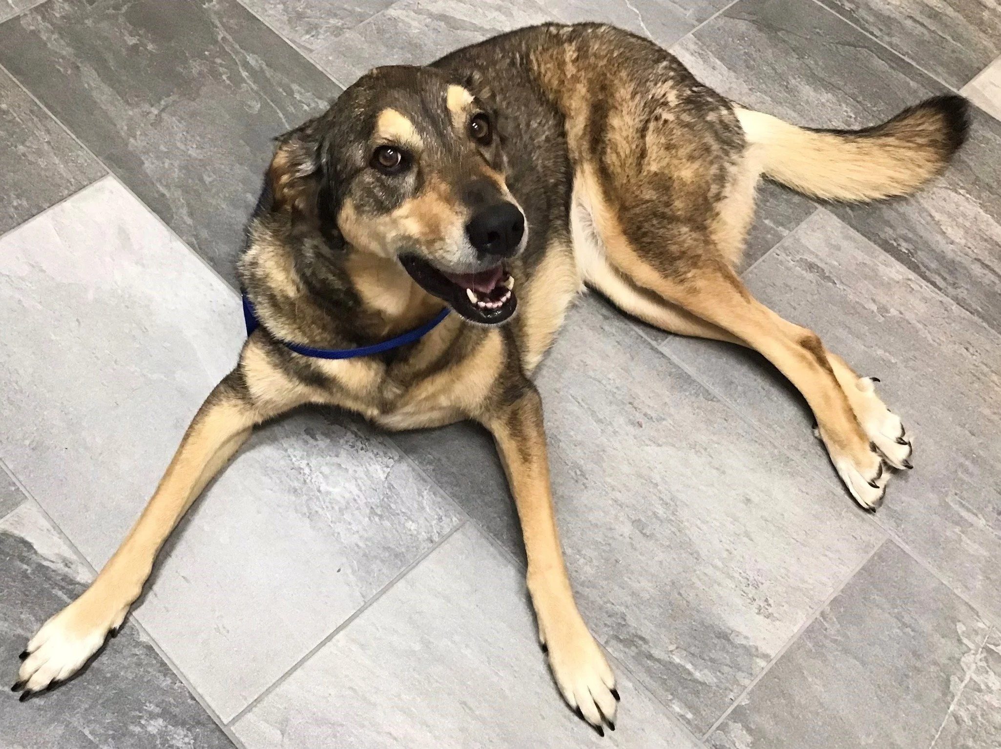
Juno, a crazy-sweet seven-year-old shepherd mix, presented to us as an emergency after she fell down while running around outside. At the time of presentation, she could not stand on her own and could not walk without falling. Her left back leg was very weak and she was dragging it.
Treatment for Juno has been supportive only, and has included rehab, protecting her from herself and bladder management.
Four months after incident on her last recheck, Juno was walking completely independently, urinating on her own, had all her facilities, and was non-painful with just a tiny limp in her left rear leg.

Juno at her recheck 20 days after initial presentation shows that she can stand on her own but she still has profound weakness in her left rear leg - she cannot properly place her left back paw properly.

A close up of Juno’s left back paw showing her paw knuckled over - she would walk on the wrong side of her paw.
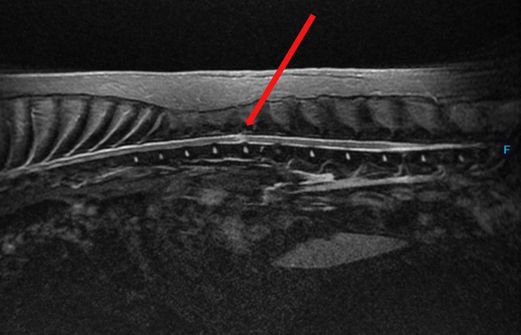
Sagittal view of Juno’s spine - MRI STIR sequence. The red arrow points toward the lesion in the middle of the spinal cord which is bright in this sequence and lies right above the T13-L1 disk space.

Axial view of Juno’s spine at the level of the T13-L1 disk space - MRI T2W sequence. The red arrow points towards the bright spot on the left side of the spinal cord (left and right projections are opposite in imaging figures).
Concerned about your pet?
If you believe your pet may be experiencing symptoms related to ANNPE, we encourage you to request an appointment with our veterinary specialists for consultation and personalized care.